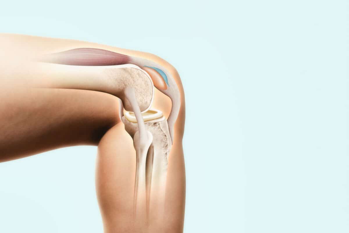
While pain may not be present, many people in their 30s already have joint damage. (© Dragana Gordic - stock.adobe.com)
In a nutshell
- Hidden knee damage is surprisingly common in young adults. MRI scans revealed that nearly two-thirds of 33-year-olds had cartilage lesions and over half had early bone growths (osteophytes), despite having little to no knee pain or symptoms.
- Weight plays a major role in early joint deterioration. Higher body mass index (BMI) was the strongest and most consistent factor linked to knee damage, more than family history, blood pressure, or uric acid levels.
- These early changes could foreshadow future osteoarthritis. Although participants felt fine now, researchers say these structural joint issues may increase the risk of developing painful, symptomatic osteoarthritis later in life.
OULU, Finland — Your knee joints might be silently deteriorating decades before you feel a twinge of pain. A new study from Finland reveals that more than half of healthy 33-year-olds already show cartilage damage in their knees, despite experiencing no symptoms.
The study, published in Osteoarthritis and Cartilage, showed that structural knee damage is remarkably common in adults just entering their thirties, even among those who feel perfectly fine. The research team found that nearly two-thirds of the young adults examined had cartilage damage, and more than half had small bone growths called osteophytes, all years or even decades before many would expect joint problems to develop.
The Finnish researchers set out to understand how common structural knee abnormalities might be in typical 30-somethings who weren’t athletes or people with previously known injuries. They examined MRI scans from 288 participants (about 61% women) with an average age of 33.7 years from the Northern Finland Birth Cohort 1986.
The study focused on individuals born in Finland’s two northernmost provinces between July 1985 and June 1986. For a subset of the cohort, researchers conducted comprehensive clinical evaluations, laboratory analyses, and knee MRIs when participants reached 33 years of age.

Participants were asked which knee troubled them more, and that knee was imaged, though most reported no significant pain or symptoms. The MRIs revealed surprising rates of structural changes that typically aren’t expected in people this young.
Over half had damage to the cartilage where the kneecap meets the thigh bone (the patellofemoral joint), and a quarter showed damage where the thigh bone meets the shin bone (the tibiofemoral joint).
The patellofemoral joint appeared particularly vulnerable. Full-thickness cartilage lesions (where damage extends through the entire thickness of the cartilage) and bone marrow lesions (areas of increased fluid in the bone) were mostly concentrated in this area.
Osteophytes were found in 51.7% of patellofemoral joints and 17.4% of tibiofemoral joints. While most were small, they are unusual in a population this young.

However, study participants generally weren’t experiencing knee problems. The average total WOMAC score (a measure of joint pain, stiffness, and function) was just 8.4 out of a possible 240, indicating minimal symptoms. Between 93% and 99% of participants reported little to no knee pain, stiffness, or functional limitations.
The factor most consistently associated with these early joint changes is body weight. The study found that body mass index (BMI) was significantly linked to increased rates and severity of knee damage, raising concerns about long-term joint health as obesity rates continue to rise worldwide.
Other factors showing associations, though less consistently, included elevated blood urate levels and systolic blood pressure. A family history of knee osteoarthritis showed some association, but wasn’t as strongly or consistently linked as body weight.
Many think that knee joint changes typically begin much later in life. The researchers cite a large cohort study that found the fraction of patients with osteoarthritis in the 18–44-year age category had increased from 6.2% in 2001 to 22.7% in 2018. They also reference the Global Burden of Disease Study 2019, which reported rising osteoarthritis incidence in the 30-44 age groups since 1990.
The researchers say that while these early knee changes aren’t necessarily painful now, they could signal future problems. Osteoarthritis, often thought of as an “old person’s disease,” may be silently forming decades earlier than previously recognized. And while many factors affecting joint health aren’t modifiable, maintaining a healthy weight appears to be one protective measure within our control.
Paper Summary
Methodology
This study examined 288 participants (176 females, 112 males) with a mean age of 33.7 years from the Northern Finland Birth Cohort 1986. Researchers conducted knee MRI scans on participants, who were asked which knee experienced more symptoms (though most were largely asymptomatic). The MRIs were evaluated using the semi-quantitative MRI Osteoarthritis Knee Score (MOAKS) system. Participants also completed comprehensive questionnaires about their medical history, lifestyle factors, and knee symptoms using the WOMAC scale. Laboratory analyses were conducted from blood samples after overnight fasting. Statistical analyses included descriptive statistics and multivariable regression models to identify associations between background/clinical parameters and MRI findings.
Results
The study found that subtle knee MRI findings were common in this young population. Cartilage lesions were identified in 56.2% of patellofemoral joints and 25.3% of tibiofemoral joints, though most were small. Osteophytes were detected in 51.7% of patellofemoral and 17.4% of tibiofemoral joints. Full-thickness cartilage lesions and bone marrow lesions were mostly located in the patellofemoral joint. Despite these structural findings, participants generally reported minimal knee symptoms, with most having no to mild pain, stiffness, or functional limitations. Of analyzed background and clinical parameters, higher BMI was most frequently associated with MRI findings, showing significant correlations with 22 out of 40 analyzed MRI parameters. Higher plasma urate levels and systolic blood pressure also showed associations with MRI findings, though less consistently than BMI.
Limitations
The study acknowledges several limitations, including the relatively infrequent occurrence of advanced MRI findings in this population, which limited statistical power for some analyses. Full-length standing radiographs for assessing axial alignment were unavailable. Despite using multivariable regression models, some residual confounding could exist. The researchers also note that with a limited sample size, some results could be based on chance. The study didn’t analyze socioeconomic status or lower limb injuries other than fractures. Finally, without longitudinal data, the study cannot determine which early joint changes will progress to symptomatic osteoarthritis over time.
Funding/Disclosures
33- 35 yThe work was supported by the Research Council of Finland (Flagship of Advanced Mathematics for Sensing Imaging and Modeling grant #359186 and grant #354692). The NFBC1986 33-35y follow-up study received financial support from the University of Oulu (Strategic funding from donations) and Oulu University Hospital (K65760). The authors declared no conflicts of interest.
Publication Information
The study titled “Structural knee MRI findings are already frequent in a general population-based birth cohort at 33 years of age” was published in Osteoarthritis and Cartilage in 2025. Authors include Antti Kemppainen, Joona Tapio, Miika T. Nieminen, Simo Saarakkala, and Mika T. Nevalainen, all affiliated with institutions in Oulu, Finland, including the University of Oulu and Oulu University Hospital.


